Siemens Prisma at Life Science MRI Facility
This page is dedicated to examples of imaging and research that has been and/or could be done here at the Purdue MRI Facility.
Anatomical Imaging
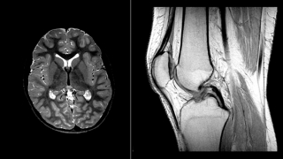
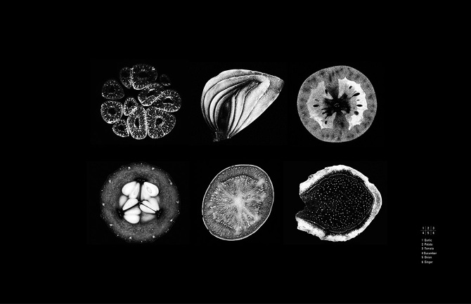
MRI (Magnetic Resonance Imaging) is a tool now commonly used in hospital and outpatient settings, used to acquire detailed imaging without the use of Ionizing Radiation. Common body parts imaged using MRI include the Brain (Left side of the Left image above) and the Knee (Right side of the Left image above). Curious what certain items look like using MRI? Examples of fruits and vegetables in an MRI environment are represented above (Right image).
Spectroscopy
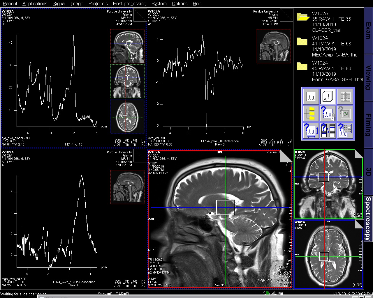
The technique of magnetic resonance spectroscopy (usually shortened to MR spectroscopy or MRS) allows tissue to be interrogated for the presence and concentration of various metabolites. Commonly, MRS is used for evaluating specific areas of the brain. However, Spectroscopy can and has been used for the evaluation of many items including the heart, liver, and other organic tissue.
Functional Imaging
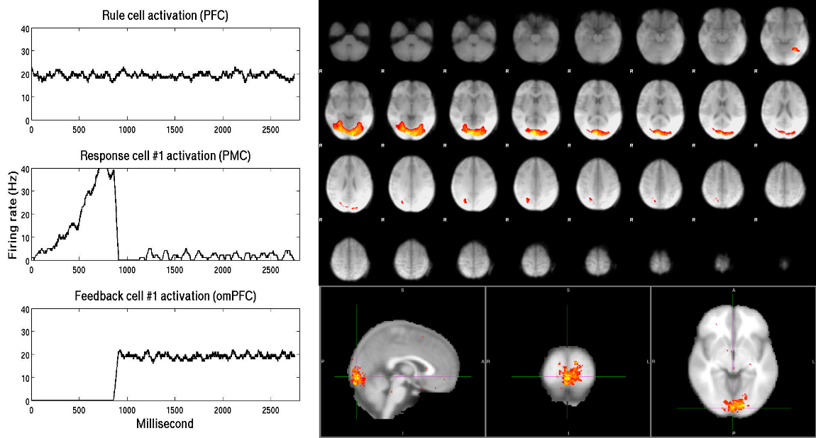
Functional magnetic resonance imaging (fMRI) measures the small changes in blood flow that occur with brain activity. It may be used to examine the brain's functional anatomy, (determine which parts of the brain are handling critical functions), evaluate the effects of stroke or other disease, or to guide brain treatment. fMRI may detect abnormalities within the brain that cannot be found with other imaging techniques. fMRI is a useful research tool that does not require any injection, but does require specific software and post-processing of raw data acquired at the MRI scanner.
Diffusion Imaging (DWI, DTI, DSI, etc.)
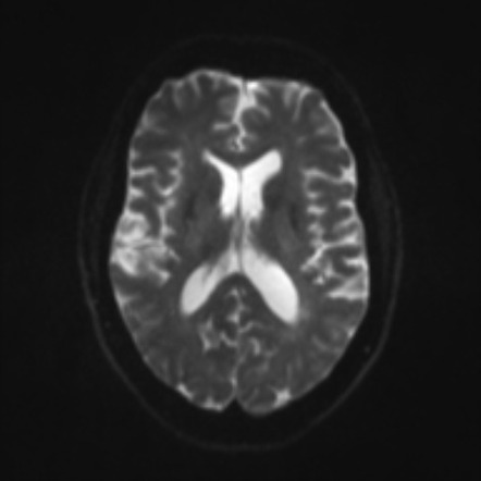
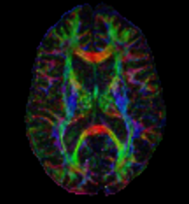
Examples of DWI on the left, DTI color maps on the right, DTI and DKI below.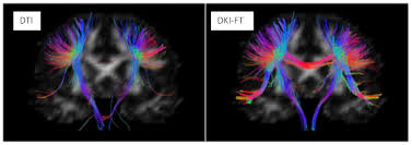
Diffusion describes the constant random motion that all molecules undergo because of their intrinsic thermal energy. Clinical diffusion weighted imaging (DWI) studies the molecular motion of water, probing its mobility on the cellular scale. The diffusion coefficient, measured in mm 2 /sec, relates the average motion, in a mean-squared sense, to the observation time, with higher values of this coefficient indicating more mobile water molecules. The apparent diffusion coefficient (ADC) characterizes water mobility observed in the clinical setting, reflecting the limitation that, in vivo, pure diffusion cannot be easily separated from other sources of water mobility, such as active transport, changes in membrane permeability, and pressure gradients.
Advances in Diffusion Imaging have yielded opportunities to evaluate and visualize not only ischemic injury and stroke, but also nerve fiber tracts. Several techniques that are being used include Diffusion Tensor Imaging (DTI), Diffusion Spectrum Imaging (DSI), and Diffusion Kurtosis Imaging (DKI).
- MRI Services
- Siemens MRI